Medical Imaging
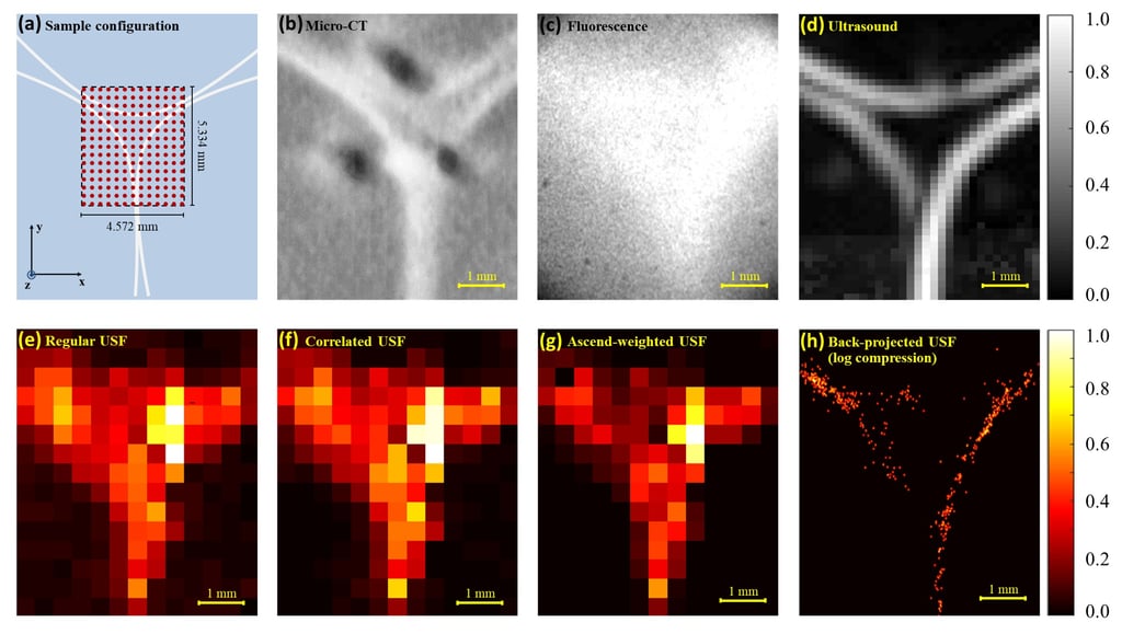
Ultrasound switchable fluorescence (USF) imaging has been proposed as a novel imaging modality which provides microscopic fluorescence imaging in centimeter-deep tissue. USF is possible to reveal many interesting phenomena such as microcirculation, tumor angiogenesis, and cancer metastasis and diagnose early-stage cancer. USF adopts a focused ultrasound (FU) to repetitively switch “on” fluorescent agents in tissue, and the USF photons scatter out and are captured by a detector. A USF image provides acoustic resolution and optical sensitivity.
In USF, there are two key components: 1) an excellent USF contrast agent; and 2) a sensitive USF imaging system. The major challenge of USF is to develop USF contrast agent with excellent USF properties and also long-term stability, bio-stability, and functionalization, as well as to develop a USF imaging system with high detection sensitivity, specificity, efficiency, and also flexible experimental operations for a desired imaging quality. In addition, in vivo USF imaging is an important step to push USF technique to future biomedical applications.
We demonstrated results by 3D co-registration with the micro-CT images and showed in vivo feasibility of USF imaging. We adopted ICG-NP as the in vivo USF contrast agents, which shows a high bio-stability in mice’s tumor as well as spleen and liver.
ULTRASOUND-SWITCHABLE-FLUORESCENCE IMAGING
Check for details in Dr. Yu's Dissertation and Paper: